Ultrasound Imaging
Acuson Sequoia 512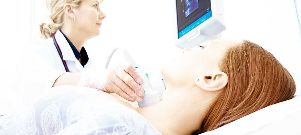
 Acuson Sequoia 512
The only ultrasound that has the coherent image formation, which is the ability to produce twice the information in half the time utilizing full echo formation without additional non-image fillers. The Sequoia also has other advanced and exclusive features that offers an unprecedented amount of new information. With this powerful tool we can now routinely see anatomy and physiology not seen with conventional ultrasound. the Acuson Sequoia 512 enables more accurate and consistent visualization of subtle tissue contrast differences and remarkable blood flow assessment.
CONTACT INFORMATION:
(772) 569-9745 OFFICE
(772) 567-6868 FAX
Medical Specialty Center
1485 37th Street, Suite 107
Vero Beach, Florida 32960
click the “+” to expand service explanation
ABDOMEN (COMPLETE)
DESCRIPTION: Examination of the liver, gallbladder, spleen, abdominal aorta, both kidneys, biliary tree and pancreas. These structures are examined thoroughly in two planes. Color Doppler is used as required to demonstrate vascularity and flow direction.
PROCEDURE EXPLAINED: The patient lies in the supine position (on his/her back) on the ultrasound table. A small amount of warmed gel is placed on the abdomen which allows proper contact between the skin and the transducer. A transducer is a device that emits ultrasonic waves through the skin and receives images back to transmit to the ultrasound screen. The transducer is placed on the skin and moved as needed to visualize each organ.
TOTAL TIME REQUIRED: Approximately 30 minutes.
ULTRASOUND - AORTA
DESCRIPTION: To demonstrate the size, depth and course of the aorta for determination of abnormality, such an aneurysm, or pathology. Pulsed Doppler and color flow images are used during this exam to determine flow and velocity.
PROCEDURE EXPLAINED: Patient lies on his/her back on the ultrasound table. Warmed gel is placed on the patient’s abdomen and the Technologist begins scanning by sliding the transducer across the abdomen to visualize the aorta. Measurements are taken throughout the length of the aorta. Color flow images are also made.
TOTAL TIME REQUIRED: Approximately 30 minutes.
ULTRASOUND – BILIARY
DESCRIPTION: To visualize the biliary ducts for dilatation or obstruction.
PROCEDURE EXPLAINED: Patient lies on his/her back on the ultrasound table. A small amount of warmed gel will be placed on the abdomen and a transducer will be scanned on the skin to produce images of the biliary system. Patient may be asked to move into different positions in order to allow the Technologist to measure the ducts.
TOTAL TIME REQUIRED: Approximately 30 minutes.
ULTRASOUND – CAROTID ARTERIES
DESCRIPTION: An examination of the carotid arteries (in the neck) to determine the presence of stenosis (narrowing), plaque (calcified particles attached to the inside walls of the arteries) or other abnormalities.
PROCEDURE EXPLAINED: Patient lies on his/her back on the ultrasound table. The Technologist places warmed gel on the neck and scans from the lowest portion of the neck to near the ear to image the carotid arteries on both sides of the neck. Doppler is also used to measure the velocity of the blood flow.
TOTAL TIME REQUIRED: Approximately one hour.
ULTRASOUND – PELVIS ENDOVAGINAL
DESCRIPTION: A study to demonstrate the pelvic organs. This exam is used routinely in combination with a transabdominal pelvic exam to further delineate the pelvic organs and subtle pathology. Specifically, endovaginal imaging should be used in first trimester pregnancies, question of ectopic pregnancy, evaluation of PID (pelvic inflammatory disease), evaluation of ovarian follicles for fertility studies, evaluation of adnexal masses and in obese patients.
PROCEDURE EXPLAINED: Patient is asked to empty her bladder in the restroom prior to the beginning of this study. The entire procedure will be explained to the patient before beginning. Patient will be given a gown or sheet for privacy. Underpants and or pantyhose must be removed. Patient will lie on her back on the ultrasound table. The transducer probe resembles a wand that has a slight bend at the end. It is approximately ¾” in diameter. This probe is covered with a latex sheath and lubricated well on the tip. Either the patient or the Technologist inserts the transducer probe into patient’s vaginal cavity. The transducer is then gently moved from left to right or down (flush against cervix or vaginal vault) to demonstrate pelvic anatomy or pathology. Color Doppler is used to demonstrate vascularity as required. Endovaginal studies allow clearer pictures in many instances because the probe can get closer to the areas of interest.
TOTAL TIME REQUIRED: Approximately 30 minutes.
ULTRASOUND – GALLBLADDER
DESCRIPTION: A study to visualize the gallbladder and biliary system for evaluation of pathology such as; stones, acute or chronic inflammation, hydrops, tumors, abscesses, polyps, etc. The pancreas and liver are reviewed for pathology also.
PROCEDURE EXPLAINED: The patient lies on his/her back on the ultrasound table. Warmed ultrasonic gel is placed on the patient’s abdomen and a transducer is placed on the skin and moved in various positions to properly visualize the organs.
TOTAL TIME REQUIRED: Approximately 30 minutes.
ULTRASOUND – INFERIOR VENA CAVA (IVC)
DESCRIPTION: The IVC is located to the right of the abdominal aorta and behind the pancreas. This study evaluates the IVC for abnormality such as dilatation, tumor, thrombus or displacement. The blood flow is documented in color duplex and Doppler images.
PROCEDURE EXPLAINED: Patient will lie on the ultrasound table on his/her back and will also be asked to turn up onto the left side during this study. Different breathing techniques are necessary to adequately visualize the IVC. The Technologist will instruct the patient when to used inhalation, exhalation and valsalva maneuver so that she may obtain unobstructed views of the IVC.
TOTAL TIME REQUIRED: Approximately 30 minutes.
ULTRASOUND – LIVER
DESCRIPTION: A study that visualizes liver parenchyma, intrahepatic structures and liver-diaphragm interface for the determination of liver size, position and the presence of intrahepatic pathology such as, liver cyst, tumors, abscesses, metastases and abnormalities of the biliary tree.
PROCEDURE EXPLAINED: The patient lies on the ultrasound table. An amount of warmed gel is placed on the abdomen and the transducer is moved over the skin in various positions to properly visualize the liver. The patient will be asked to hold their breath at times and ultrasound images will be made.
TOTAL TIME REQUIRED: Approximately 30 minutes.
ULTRASOUND – LOWER ABDOMEN / PELVIS
DESCRIPTION: An evaluation of the organs (colon, appendix, small bowel, lymph nodes, urinary bladder) and or surrounding area. Endovaginal ultrasound and color duplex and Doppler are performed if necessary.
PROCEDURE EXPLAINED:Patient is asked to lie on his/her back on the ultrasound table. Warmed gel is placed on the lower abdomen and the Technologist scans by sliding the transducer across the abdomen. Images of the specific area of interest are made. Measurements of the full bladder are taken and the patient will be allowed to empty his/her bladder in the restroom. Images are taken after the patient voids to measure the remaining urine in the bladder.
TOTAL TIME REQUIRED: Depending upon degree of difficulty, 30 minutes to one hour.
ULTRASOUND – PANCREAS
DESCRIPTION: A study to visualize the pancreas and surrounding area; to aid in the diagnosis of conditions such as pancreatitis, pseudocyst and cancer.
PROCEDURE EXPLAINED: Patient lies on his/her back on the ultrasound table. A small amount of warmed gel will be put on the skin. A transducer will be used to scan along the skin surface to image the pancreas. Patient may be asked to move into different positions for better visualization.
TOTAL TIME REQUIRED: Approximately 30 minutes.
ULTRASOUND – PELVIS TRANSABDOMINAL
DESCRIPTION: A study to demonstrate the pelvic organs
PROCEDURE EXPLAINED: Patient is asked to lower waistband of pants or skirt to a level just above the pubis. A towel is used to protect clothing from gel. Warmed gel is placed on the lower abdomen and the Technologist examines the pelvis by scanning the area with the transducer. Images are made. Patient is then allowed to empty bladder in restroom.
TOTAL TIME REQUIRED: Approximately 30 minutes.
ULTRASOUND – PREGNANCY DETERMINATION and FIRST TRIMESTER AND/OR PREGNANCY R/O ECTOPIC
DESCRIPTION: An examination of the gravid uterus via transabdominal and endovaginal (up to approximately ten weeks) methods. Specific areas demonstrated dependent upon the gestional age. Also performed to document any pathology; i.e., in cases of fetal demise, impending abortion, incomplete abortion, ectopic pregnancy, etc.
PROCEDURE EXPLAINED: Patient lies on her back on the ultrasound table. Warmed gel is put on the abdomen and the Technologist scans by placing the transducer on the skin and moving it across the abdomen to obtain images of the uterus. This is called the ‘transabdominal’ portion of the exam. When the Technologist has completed this portion of the study, the patient is allowed to go to the restroom to empty her bladder. She then returns to the ultrasound room and the second part of the exam is performed by using an ‘endovaginal probe’. This probe resembles a wand with a slight bend at the end. It is used to examine the uterus and surrounding tissues more closely via insertion of the probe into the vaginal cavity. The probe is properly maintained by decontamination in Metricide and always covered with a latex sheath during usage. The endovaginal method of scanning the uterus is used in the first trimester of pregnancy up until approximately ten weeks as deemed necessary.
TOTAL TIME REQUIRED: Approximately one hour.
ULTRASOUND – RENAL/ URINARY TRACT
DESCRIPTION: A study to demonstrate the size, shape and characteristic tissue of the kidneys, urinary bladder and adjacent structures. Evaluate for pathology such as cysts, masses, stones and other abnormalities.
PROCEDURE EXPLAINED: Patient lies on his/her back on the table and a small amount of warmed gel is put on the skin. The transducer is scanned over the skin to obtain pictures of each kidney. Patient will be asked to turn up on one side and then the other to obtain the best views of the kidneys. Ultrasound images of the urinary bladder will also be taken. The patient will then be asked to empty his/her bladder in the restroom.
TOTAL TIME REQUIRED: Approximately 30 minutes.
ULTRASOUND – RETROPERITONEUM
DESCRIPTION: The peritoneum is, very basically, the abdominal lining within which all organs are contained. The retroperitoneum is the area behind or external to the peritoneum. This exam is performed specifically to evaluate that area for displacement of organs, tumors, enlarged lymph nodes and or ascites (fluid).
PROCEDURE EXPLAINED: The patient will begin the exam on his/her back on the Ultrasound table. An amount of warmed gel will be placed on the abdomen and the Technologist will begin her evaluation by sliding the transducer on the skin over several areas of the abdomen and take images. Patient may be asked to change positions, allowing for better visualization of certain areas.
TOTAL TIME REQUIRED:Depending upon degree of difficulty, allow 30 minutes to 1 hour.
ULTRASOUND – SCROTAL
DESCRIPTION: An examination to determine the presence of varices, tumor or other pathology or abnormality.
PROCEDURE EXPLAINED: Patient will lie on his back on the ultrasound table. Technologist will provide toweling for privacy covering the genitals. Warmed gel will be placed on the scrotum and a transducer scanned over the areas to be imaged for abnormality. Color Doppler and spectral waveform analysis may be used to determine vascularity.
TOTAL TIME REQUIRED: Approximately 45 minutes.
ULTRASOUND – SPLEEN
DESCRIPTION: The spleen lies on the left side of the abdomen. This study is to demonstrate the size and characteristics of the spleen and to aid in the diagnosis of enlargement, hematoma (a collection of blood), lymphoma, etc.
PROCEDURE EXPLAINED: The patient lies on his/her back on the ultrasound table. Warmed gel is placed on the patient’s abdomen and the Technologist examines the spleen by sliding the transducer across the abdomen. Patient may be asked to turn up onto his right side for better visualization. Color flow images and pulse Doppler may be used to demonstrate the blood vessels.
TOTAL TIME REQUIRED: Approximately 30 minutes.
ULTRASOUND – THYROID
DESCRIPTION: A study that visualizes the lobes of the thyroid gland for determination of size, position and the presence of any pathology such as cysts, tumors and other abnormalities.
PROCEDURE EXPLAINED: The patient lies on his/her back on the ultrasound table. Pre-warmed gel is placed on the neck and the technologist scans by moving the transducer along the skin in various positions until she has taken pictures of all areas of the thyroid. Color Doppler and spectral waveform analysis may be used if needed.
TOTAL TIME REQUIRED: Approximately 30 minutes.